While Poisoning Others...
take care not to poison yourself

Introductory note: my wife and my daughter have sometimes told me that my essays get too science-y, and that my readers might be put off by this. They are probably right, but geez, I spent my life in science and science is how I approach the external world. I may not be the best judge of science-yness, still, I am going to risk posting this rather science-y essay in the hopes that many of my readers will bear with me, and thereby get a glimpse of a few of life’s hidden marvels.
As even middle-schoolers know, arthropods have an exoskeleton, a secreted outer covering that is both a skeleton giving form to their bodies, but also a barrier to nasty things from the outer world. What middle-schoolers probably don’t know is that to make such an exoskeleton, the animal’s epidermis (or hypodermis) is a chemical factory that makes a wide range of chemicals needed for making that exoskeleton. Moreover, like many human factories, once you’ve got the building and the machinery, you can make any number of products as well as switching among them as the situation demands.
The basic ingredients needed to build an exoskeleton are (1) a protein gel that can be hardened into shapes, a sort of casting material; (2) the chemicals needed to do the hardening that incidentally give color; (3) waxy stuff for coating the exoskeleton to reduce water loss. Thing is, several of these needed chemicals are toxic even to the cells that synthesize them, so the cells have evolved a work-around by synthesizing the non-toxic precursors of the cuticle inside the cell, then excreting them to the outside along with enzymes that complete the final chemical step resulting in the exoskeleton. Once hardened, these materials are no longer toxic. In this way, the toxins are made outside the cell, and the cell is safe from its effects.
This system is a “pre-adaptation” for the heavier artillery that is the subject of this essay. Many biologists huff at the term “pre-adaptation” because it seems to imply intent on the part of evolution, and in the words of Monty Python, intent is not “A-1 Theory With Our Lot.” To be clear, I don’t mean to imply intent, I am merely emphasizing (yet once again) that evolution always molds the clay already in its hand. In the present case, the “there” is an epidermal factory capable of manufacturing and exporting a wide range of chemicals with a few twitches of the machinery.
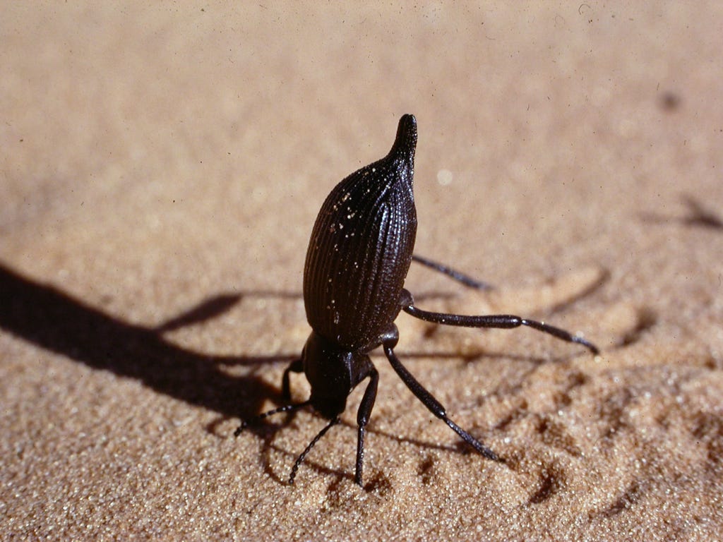
Back when I was a mere scientific puppy, I heard a very exciting talk by Thomas Eisner that lead me to undertake more than a decade of research on the chemical defensive system of tenebrionid beetles, aka darkling ground beetles. The more spectacular ones, such as Eleodes armata, can spray their noxious secretion for almost a meter, god help you if it gets in your eyes (see below). What’s more, by doing a headstand, these beetles warn you that they are going to squirt if you persist (see image at the top). Eleodes is one of about 1500 species of tenebrionids in North America, and 12,000 in the world.
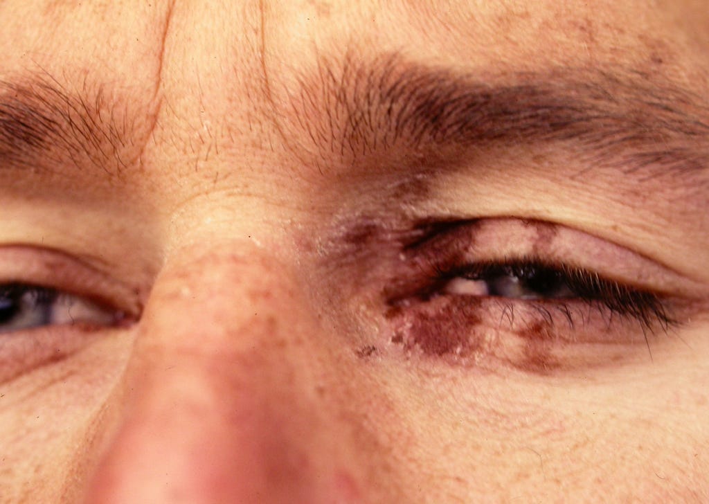
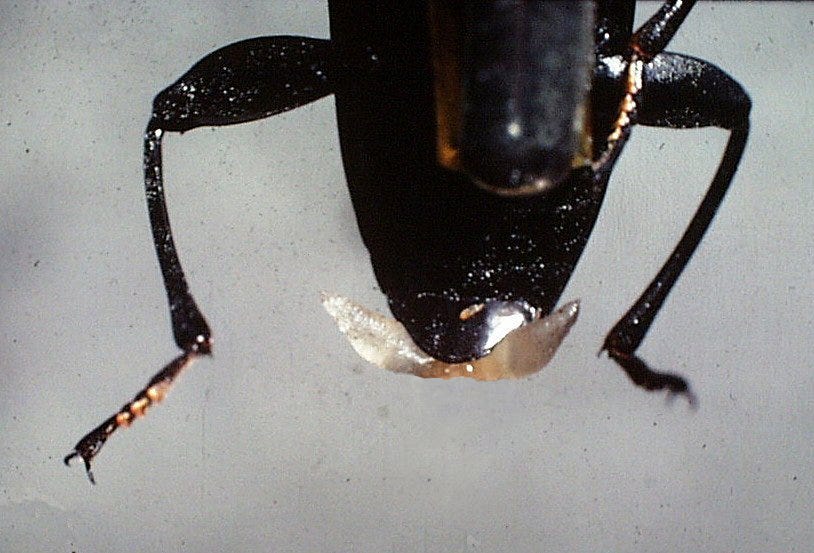
But the tenebrionid ancestor of Eleodes didn’t start with such heavy artillery. For some reason, that ancestor had a fold of soft, intersegmental membrane at the tip of its abdomen, a pocket into which it began to excrete an excess of the same quinones that had tanned its cuticle after a molt. Quinones are oxidizers, tanning agents and strong irritants with a sharp odor and a repellent nature. When this ancestral tenebrionid was occasionally attacked by a predator, it almost incidentally exposed this irritant-coated intersegmental membrane in its struggles, perhaps slightly increasing its survival in proportion to the amount of noxious quinone secretion and size of the pockets. This “starter” condition is still represented today in the common mealworm beetle and its close relatives. Hold one of these beetles between your fingers, and it will evert these small quinone reservoirs as in the image below. The secretion bites the nose and causes brown spots on the skin (it’s a tanning agent!). Release the beetle and the glands are retracted once more.
But as the hundreds of species of tenebrionid beetles evolved from this ancestor with its simple pocket of quinones, the defensive value increased through increased reservoir size, gland size and quinone volume. I set out to map these changes on the evolution of tenebrionids by analyzing the defensive chemicals, gland morphology and behavior of over 150 species. I collected the emitted secretions when the beetles were handled and threatened, separated and analyzed the chemicals with a range of physical and chemical methods, and observed the defensive behavior.
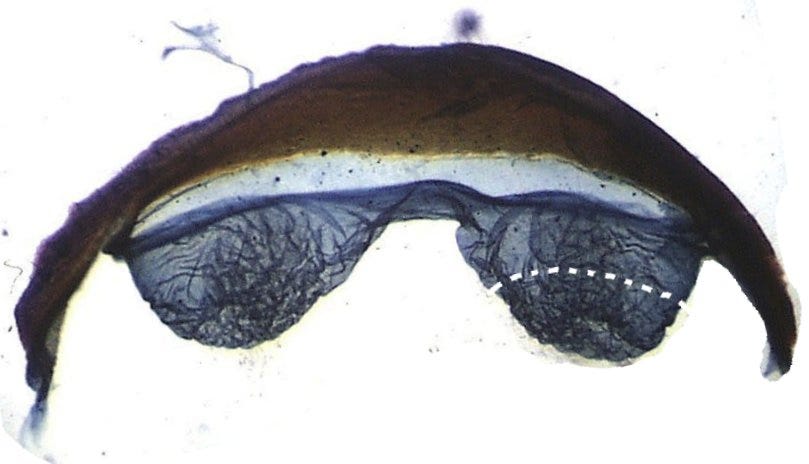
I studied the gland morphology by fresh dissection as well as in preparations made from dead, dried beetles, including long-dead museum specimens. This is one of the wonderful things about studying insect morphology—- the gland reservoirs are part of the exoskeleton made of cuticle and therefore remain behind intact after all the soft tissue has been dissolved in strong base such as sodium hydroxide (Drano!). It’s sort of cook and look. Even tiny details and fine structures remain intact. The gland reservoirs can be freed from the ventral abdomen as in the picture below, and mounted on slides for detailed study.
Below is such a preparation of the simple glands of the mealworm beetle, so similar to what the ancestral gland must have looked like. Why the mealworm beetle didn’t evolve a fancier gland in its millions of years on earth is a bit of a mystery.
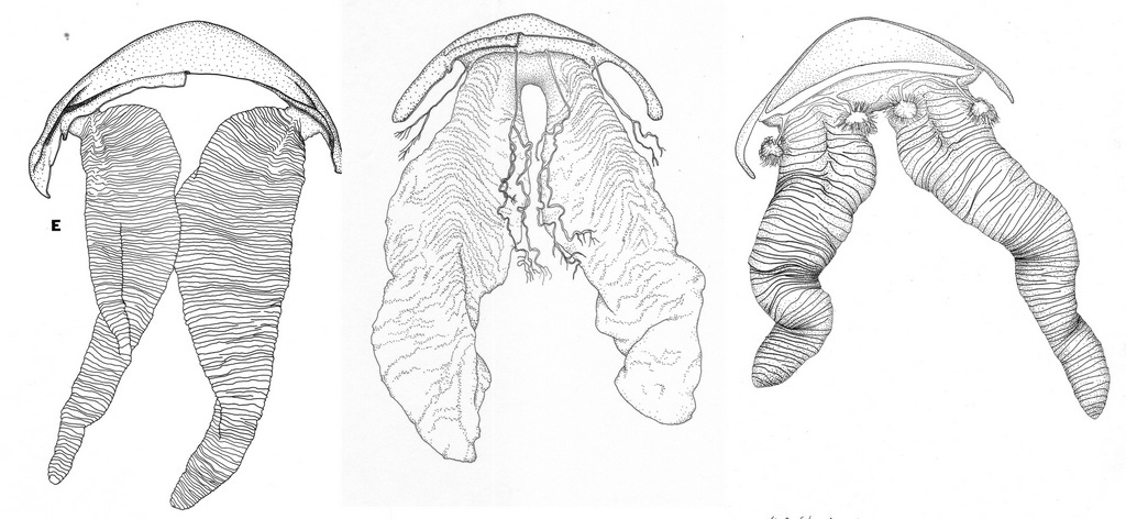
However, that is not what happened during the evolution of most other tenebrionid species, many of which got very large and fancy glands indeed, with reservoirs sometimes as long as the whole abdomen, and plenty of secretion to hose down a careless attacker. In addition, most species also added simple, linear hydrocarbons of nine to fifteen carbons (basically paint thinner) to the mixture to produce a solution with increased irritating quality, decreased viscosity, and increased ability to wet/penetrate skin and cuticle. These noxious liquids are stored safely in the reservoir through the chemical-physical properties of both the solution and the reservoir. So once this mephitic mixture is in the reservoir, it is sort of harmless to the beetle, a cannon round waiting to be fired.
Making this stuff faces the same problem that making the exoskeleton did— the reagents are toxic to living tissue, including the cells that make them. Part of my project involved detailed light and electron microscopy of the glandular cells. In the simplest case, for example in the mealworm beetle, the cells are scattered about the inside surface of the reservoir. Two types of cells are present on all tenebrionid defensive glands, and all have the same ultra-structure under the electron microscope, but vary in their distribution and drainage into the reservoir. Along the way, electron microscopy reveals that life is beautifully structured at all levels, from the organism to the cell, to the molecule.
Speaking of electron microscopy, it always surprised me that you can pickle tissue in really toxic stuff, precipitate all their biomolecules into shoe leather, dose them with the deadly heavy metal oxide of osmium, embed this micro-carcass in plastic, slice off sections as thin as a wavelength of light that you float on water, and then pick these up on a super-fine copper grid, pop this into the high vacuum of an electron microscope, and blast it with an intense electron beam. On a fluorescent screen you then see the shadow created by how much the variable density of the specimen impedes the electron beam.
And yet, the images, as near miraculous as they are, tell us a great deal about the structure of cells at a magnification far greater than achievable by light microscopes. Of course, my part of this miracle was simply to provide the dissected gland tissue and pickle it. All the rest was done by salaried experts in electron microscopy on a super-expensive instrument with a super-expensive maintenance contract. But what a thrill to get the images of my beetles at sub-micron detail, full of wonder and beauty, and to interpret how these revealed the three-dimensional structure of the gland cells. Admittedly, I wasn’t the first to reveal that this kind of structure occurred widely in insect “exocrine” (secreting to the outside) glands, but of course, adding more examples helps confirm the generality of this structure.
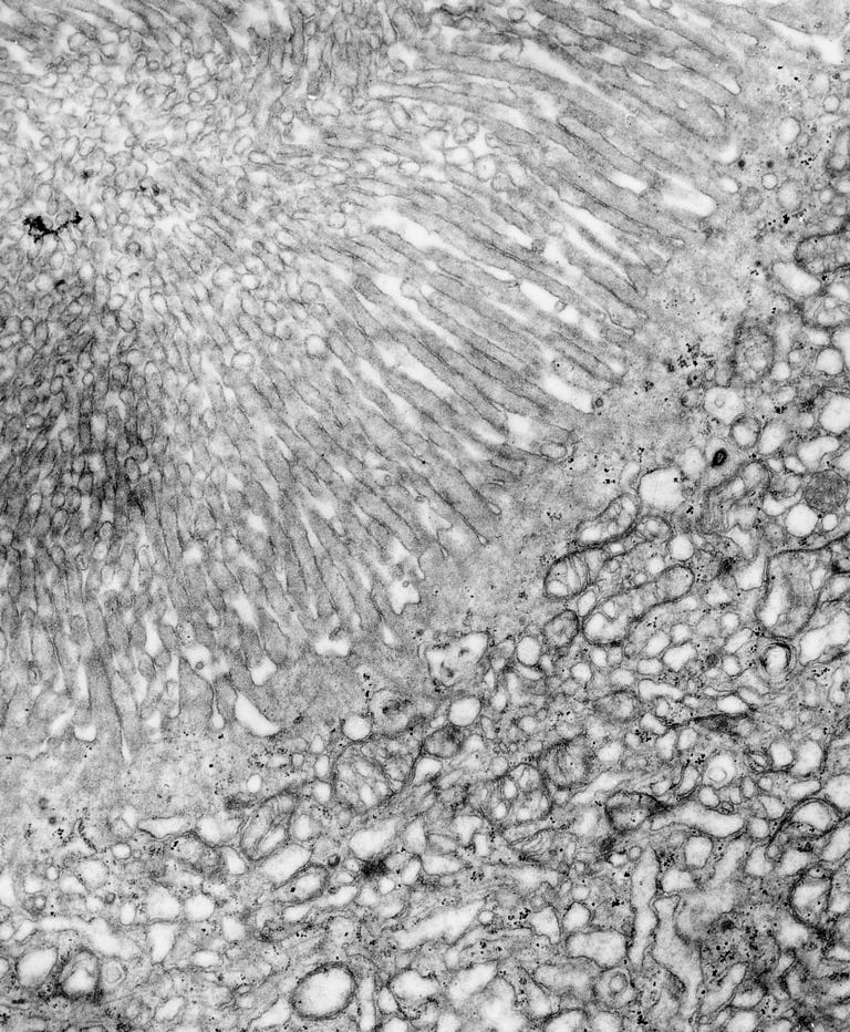
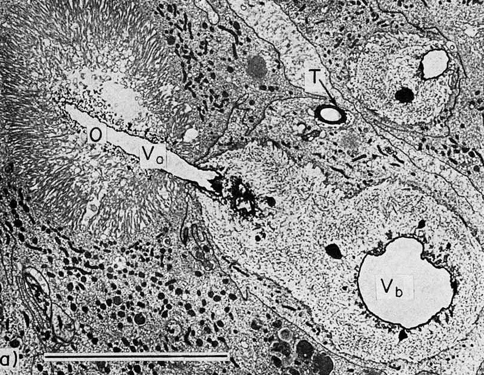
Light and electron microscopy confirmed that each of the two cell types (brilliantly named, type 1 and 2) has a central cavity (vesicle), and that of type 2 is filled with extensions of the cell membrane into finger-like projections called microvilli (an aside: in response to the test question: define microvilli, one student wrote “small rental apartments on the Italian Riviera.” I gave him full credit). Microvilli enormously increase the surface area of the cell membrane and are reliably and consistently associated with cells whose job is to exchange a lot of stuff across cell membranes, including secretory cells that make a variety of defensive secretions. In tenebrionid beetles, one type of cell secretes quinones and the other one hydrocarbons.
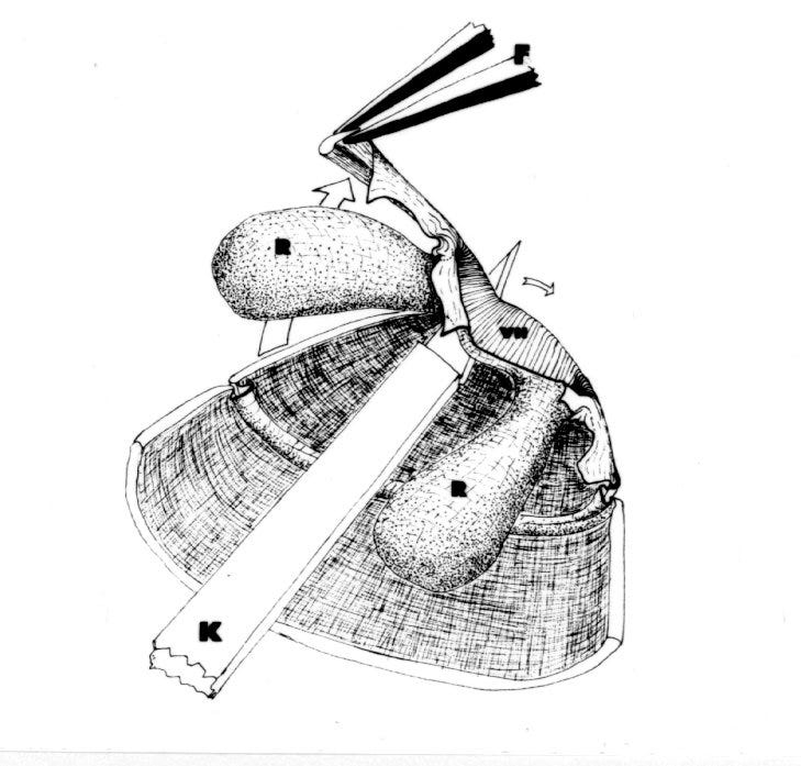
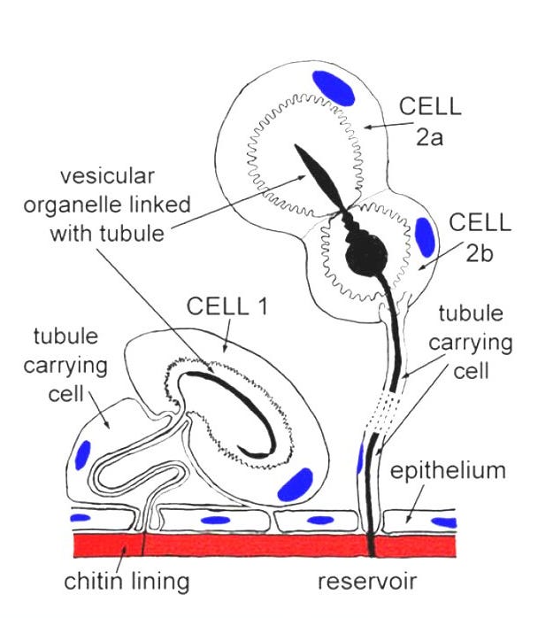
In the center of each vesicle is a cuticular structure that is continuous with the reservoir wall, essentially an extension of the reservoir wall into the heart of the secretory cells. This cuticular extension emanates from the reservoir via a cuticular duct secreted by an associated cell giving a three-cell unit (type 2), and a two-cell unit (type 1) as in the schematic below.
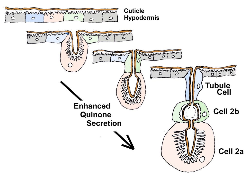
I suggested at the beginning of this essay that secretion of cuticle by the hypodermis was the pre-adaptation from which the defensive glands evolved, including at the cell level. The schematic below shows how I think the type 2 unit evolved from the elaboration of ordinary hypodermal cells that greatly increased their ability to make some of the chemicals needed to make cuticle, but were now exaggerated and “repurposed” for a new function, defense. Type 1 cells would be similar in their evolution, but are not shown.
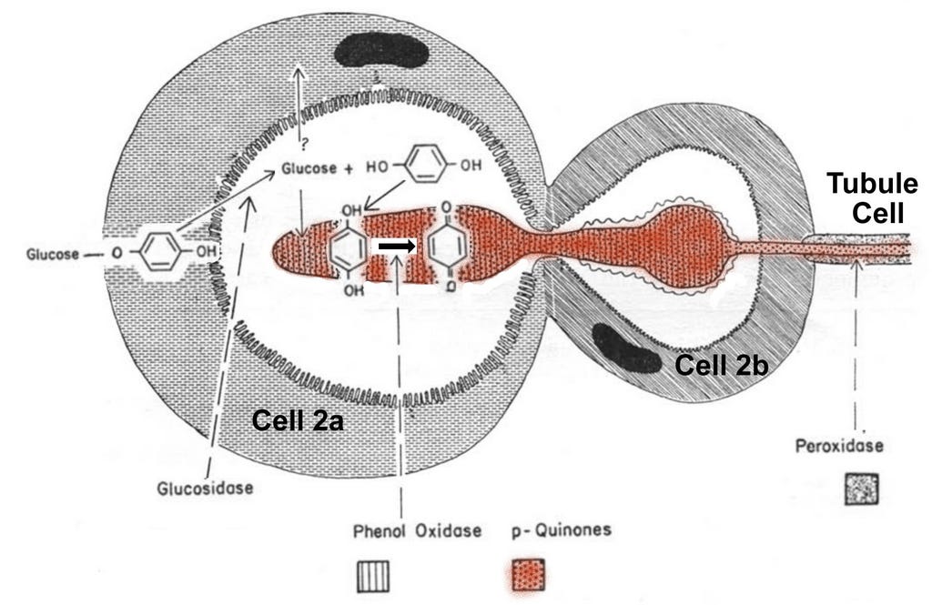
So how do these little cellular factories work, and how do they avoid poisoning themselves? In the late 1960s, George Happ did a series of histochemical and microscopic studies of two tenebrionid species (Eleodes and Tribolium) to answer this question by localizing the precursors and their products to different parts of the cells (J. Insect Physio. 14: 1821 (1968)). Thus, in the cytoplasm of the 2a cell, he detected only the precursor of quinone, hydroquinone bonded to a glucose molecule, which rendered it both harmless and water soluble. In the vesicle, he found the enzyme glucosidase, plus free glucose and free hydroquinone because the enzyme had cleaved the bond between hydroquinone and glucose. Inside the central cuticular organelle, the enzyme phenol oxidase was busily oxidizing the hydroquinone into the irritant, toxic quinones. As this stream moved toward the reservoir, another enzyme (peroxidase) continued the oxidation of the quinone precursors even in the tubules that finally emptied into the reservoir. Through similar methods, George showed that the hydrocarbons were secreted by the type 1 cell, again with the pattern of carrying out the final step outside the cell. The stored secretion in the reservoirs confirms these chemical steps, for it consists of an aqueous phase that contains the former blocking molecule glucose and the enzyme proteins, and a separate organic phase with the toxic quinone/hydrocarbon solution, nasty things dissolved in paint thinner.
So there you have it, the rather science-y story of how insects produce toxins to which their own cells are not immune. Solving how they do this engaged a lot of different procedures and skills, not all of them mine—- dissection, behavioral observation, chemical analysis and identification, electron microscopy, and histochemistry. In the classical sequence of most biological studies, these moved from level to level, from gross morphology, to fine structure, to cells, to cellular organelles, and finally to chemicals and enzymes. Together, these revealed the secret that evolution has discovered many times across the animal kingdom for many different secretory systems—- export the harmless precursors plus the enzyme and execute the final step outside the cell, and do so by modifying a capacity and machinery that is already present.
Postscript: This essay brings to mind a poem, Terrence, This is Stupid Stuff, by A.E. Houseman, the last verse of which is—-
There was a king reigned in the East:
There, when kings will sit to feast,
They get their fill before they think
With poisoned meat and poisoned drink.
He gathered all that springs to birth
From the many-venomed earth;
First a little, thence to more,
He sampled all her killing store;
And easy, smiling, seasoned sound,
Sate the king when healths went round.
They put arsenic in his meat
And stared aghast to watch him eat;
They poured strychnine in his cup
And shook to see him drink it up:
They shook, they stared as white's their shirt:
Them it was their poison hurt.
-I tell the tale that I heard told.
Mithridates, he died old.
Keep writing, I always enjoy reading. Let me assure you, I would never pick up such a vicious looking creature, no matter how small!
The science, the detail, the search for understanding and explanation, the enthusiasm are amazing and inspiring. Though I don't understand the complexities, you continue to expand my appreciate of evolution and your work.