

On Being Transparent
Insect anatomy made easy

Many small animals are transparent to varying degrees, and to a biologist like me, it is always pleasing to see their insides at work. Many freshwater worms and larval insects reveal their constituent parts, and with some basic knowledge, it is fun to identify their various organs and organ systems, and to marvel at their structure and function at this tiny scale. Among these “animals without secrets”, my favorite is the nymph of a dragonfly. I wrote an essay on the amazing gill that has resulted from a marriage of the tracheal (respiratory) system with part of the digestive system. But there is so much more to see and I offer a guided tour of this lovely insect, a glimpse into the details of the parts that make the whole work. There are, of course, parts that are “not open to the public,” and there are also adult insect parts that are not present, chiefly gonads and their associated reproductive structures. The adult wings are represented only by the small wing buds that will eventually grow into the wings. Here is a portrait of the star of this essay.
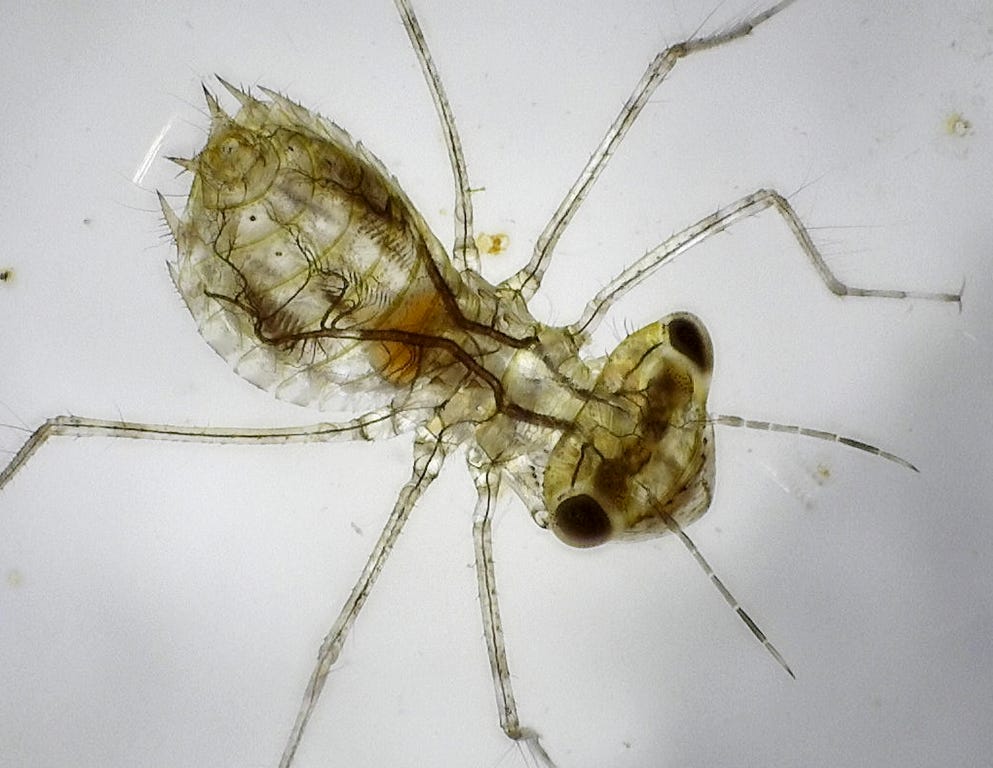
Let’s start the tour with the most prominent system in the image above —-a network of dark, branching tubes that make up the air-filled tracheal (respiratory) system. In land-based insects, this system would open to the atmosphere through spiracles on the sides of most segments, but in this aquatic nymph, the spiracles are closed and a gill is fashioned from the union of highly divided tracheae and the highly-folded wall of the hindgut. I described this gill, its functions, and its evolution in another essay. Gases that are exchanged in this gill are distributed by the huge, longitudinal trunk tracheae that run much of the dorsal length of the nymph, with pigmented branches serving all regions of the body, including the full length of all the legs and antennae. Tracheae branch and re-branch until they end in transparent microscopic branches in the vicinity of every body cell as they consume oxygen and emit carbon dioxide. I have no idea why the tracheae are pigmented. Maybe just so we can see them more easily? And why not? Everything else about this creature seems designed to be seen.
The head is like a glass capsule into which we can peer. The large, dark compound eyes are unlike those of the bee or fly whose faceted outer surface is all you see. In contrast, the nymph’s eye is largely transparent, revealing its construction from sheaves of elongate ommatidia and a layer of dark pigment. I don’t know whether the pigment is the photoreceptive pigment or the shielding pigment to restrict the light to each ommatidium. The output of the ommatidia travels down the optic nerve to the prominent optic lobe, which is essentially part of the brain, and undoubtedly has a role in image formation. Don’t ask me what the dark speckles on the optic lobe are, because I don’t know. From the size of the eye and optic lobe, you can guess that the nymph is a very visual creature, sensing the movement and location of any creature in front of its face, and then grabbing it with its protrusible labium (which I describe in another essay). Think of the nymph as a praying mantis, only underwater, but just as fast, and with the same lie-in-wait strategy.
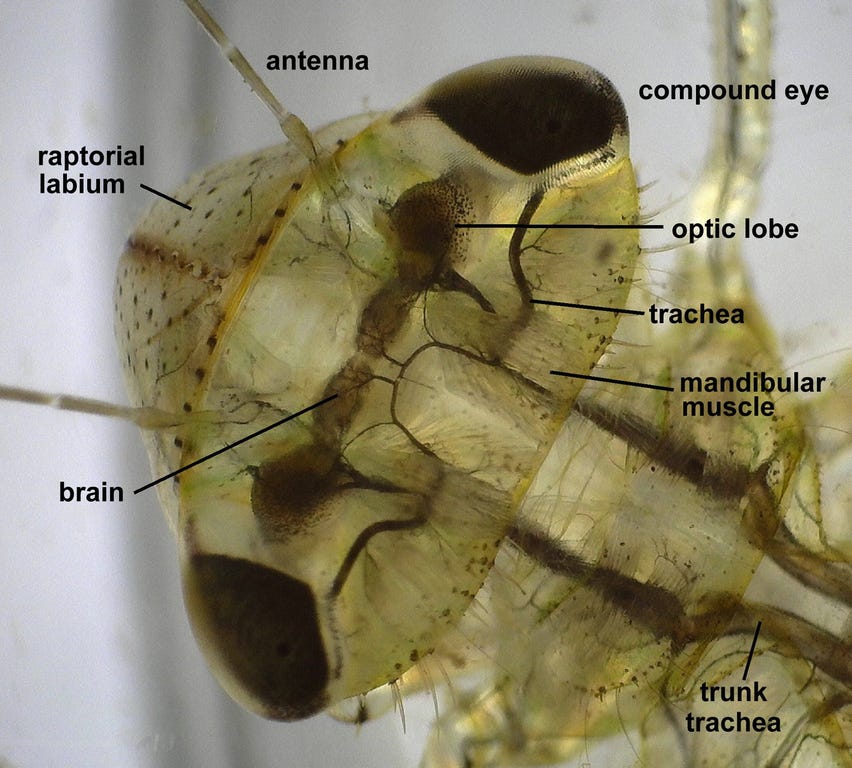
The central bilobed brain, as in all insects, is not very large because it is not the counterpart of the vertebrate brain with its central control. The insect brain is connected to a string of ganglia on a ventral nerve cord, one ganglion per segment, and much control is delegated to these ganglia. The ventral nerve cord is not visible in the image above, but runs around the esophagus and then the length of the animal.
There is much more to see within the glass-like head. The dark, pigmented tubes that branch and rebranch are the air-filled tracheal system mentioned above. In the image below, you can see branching and rebranching to serve the brain and the optic lobe, and the large trachea that keeps the eye happy. To judge from the size of the tracheae, the tissues they serve are metabolically very active, especially the eye and the brain.
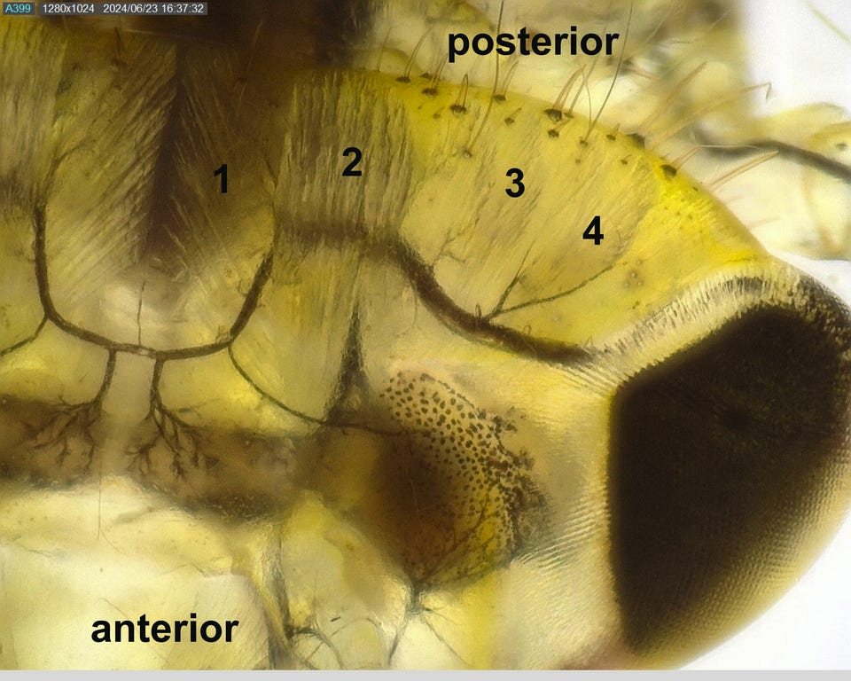
The head contains the mouthparts, and these require a lot of muscle to move. Muscles that close the mouthparts to bite or chew are very large, as they require a lot of force, and are inserted on the inside of the head capsule. Four such muscles running at different angles are visible in the image above (numbered). Their attachments to the mouth appendage are below the brain and optic lobe, and are thus not visible from above. Muscles that open the mouthparts are smaller, as they don’t need a lot of force. I don’t know which closer muscle serves which particular mouthpart, but they probably stack in the same order as the mouthparts— mandibles, maxillae, labium.

Continuing back from the head, the central nervous system consists of a double, ventral nerve cord with a ganglion in each segment and a brain that is dorsal (as we saw) to the esophagus. The first ganglion behind the brain is in the head and is a composite of (at least) three ganglia that control the mouthparts (part of whose muscles we saw in the previous image) and receive sensory input from the sensory palps and antennae. Moving from front to back, there is a ganglion for every segment, though some may be fused into a single structure. Five of the eight abdominal ganglia are labeled in the image above. The lateral branches that innervate each segment are easily visible, as is the double ventral nerve cord. Sensory input travels to the central nervous system to be processed and (if needed) acted upon.
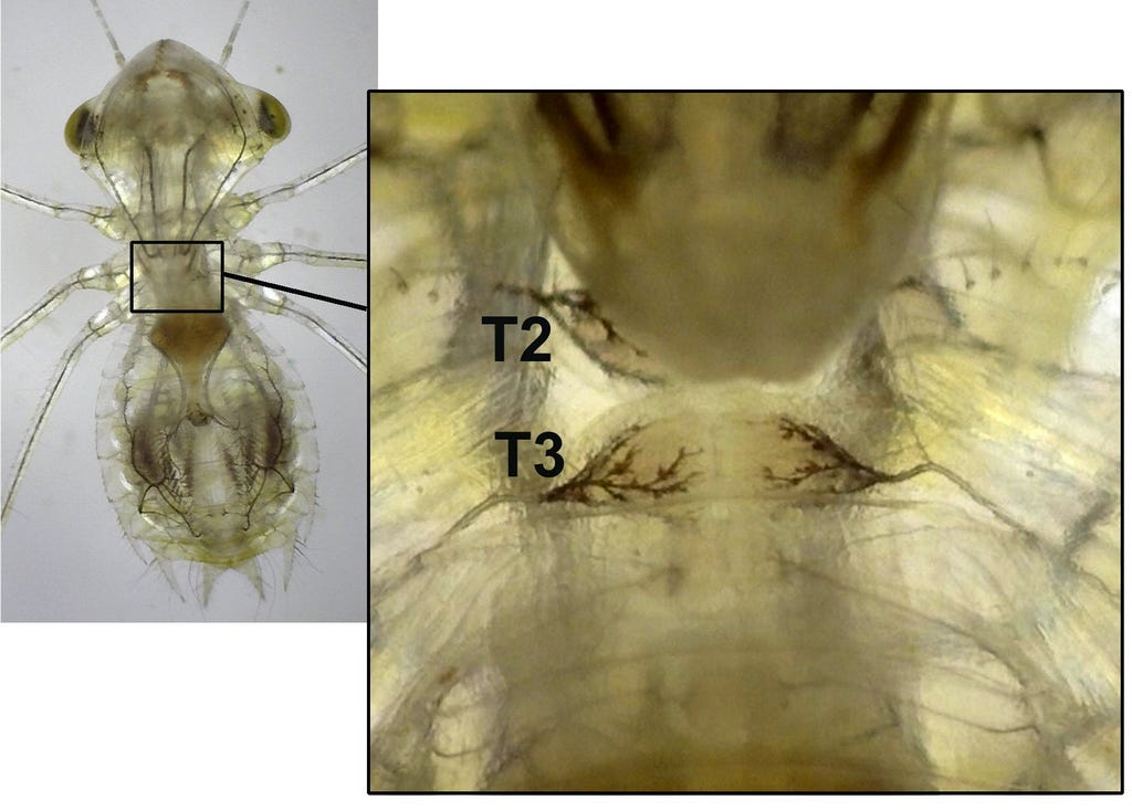
Each of the three ganglia in the thorax controls a pair of legs and receives input from them. In the ventral view below, the huge labial apparatus covers the first thoracic ganglion, part of the second and most of the face. Still, because they are well-supplied with dark, branching tracheae, it is clear that these thoracic ganglia are all larger and metabolically more active . You can also see the transparent muscles of the thorax and legs.
As mammals, we tend to think of a circulatory system with red, oxygen-carrying blood confined to a system of arteries, capillaries, and veins. But, as I wrote in an earlier post, the main function of insect circulatory systems is not the transport of gases. Insect have an open circulatory system in which the blood bathes the tissues directly, transporting nutrients, hormones, waste products, and so on. The space through which the blood flows is called the hemocoel or blood space. Insect blood is therefore colorless, and the cells it contains do not carry oxygen. Still, the blood must be circulated, and to that end, insects have the analog of the vertebrate heart, a pulsatile dorsal blood vessel that resides mostly in the abdomen, and that has lateral openings into the hemocoel from which it draws in the hemolymph. The contractions propels the hemolymph forward to the head, where it is dumped into the hemocoel just behind the brain to make its way back to the body and the legs for another round. In the video below, you can see the vessel’s chambers, one per abdominal segment with no-return valves between them. The pumping rate is quite variable, presumably responding to demand, but the waves of contraction almost always move toward the head.
The blood may be colorless, but it does contain large numbers of cells that can be made visible with backlighting at high magnification. Insect circulation is often described as rather sluggish, but this is probably vertebrate chauvinism. The video below shows torrents of hemolymph rushing around the body in organized streams. The hemocoel is divided into dorsal, ventral and visceral zones by septa, but these septa are not visible in the video.
Getting hemolymph down to the ends of those six long appendages is obviously a challenge, but as ancestral insects all had six legs and many have wings as well, it is a problem that they solved a long time ago. The length of each appendage is divided by a longitudinal septum into an outgoing and a return flow channel. At high magnification, such outward and returning blood streams become easily visible in the video.
Because the hemolymph bathes all the tissues directly, it distributes metabolites as well as receives cellular waste products. The waste products are excreted by a system of transparent tubes called the Malphigian tubules. They are the analog of our kidneys, and work on similar principles. The tubules snake sinuously throughout the hemocoel in the spaces between tissues. Thus, the insect’s “kidney tubules” are distributed throughout the body, rather than concentrated in an organ serviced by a branch of the circulatory system, as in vertebrates. The tubules open into the gut at the junction of the mid and hindgut, and look for all the world like the head of Medusa whose hair is a mass of writhing snakes, as you will see in the video above. At their outer ends, hemolymph (minus the proteins) is taken up by the Malphigian tubules, and as this fluid travels down the tubes, all the “useful” items are resorbed, and the final waste product is voided into the hindgut, and from there, through the anus.
Insect muscles, like all muscles, can only contract and shorten. Their return to their original length requires the contraction of an antagonistic muscle attached to the moving structure on the other side of a fulcrum. Muscles thus mostly work in pairs—- an adductor and an abductor. You can see this operation in your own limbs. Like most other things in our little nymph, muscles are transparent, visible mainly by their differences in refracting light. The image below shows the paired muscles that operate the two hind legs, as well as the ganglia that control them.
We’ve already seen the major muscles in the head that operate the mouthparts. The video below shows the structure and operation of a number of muscles, including the intersegmental body wall muscles that run the length of each segment, attaching and originating on the intersegmental membranes. These allow the abdomen to be compressed and may play a role in shooting out the labial, raptorial mask.
Like the adult dragonfly, this nymph is a voracious predator, but unlike the aerial hunter adult, the nymph is a lie-in-wait predator, using a highly modified labium (the hindmost mouth appendage) to snatch small prey that stray across its visual field. This raptorial labium deserves an essay of its own.
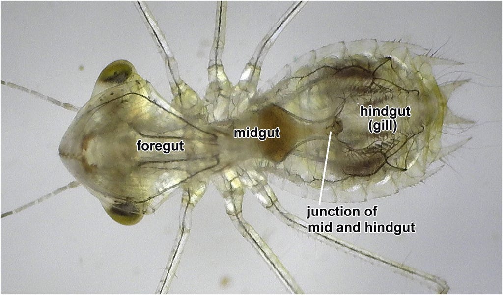
A final obvious system is the gut, which in insects consists of a cuticle-lined foregut, a digestive midgut, and a cuticle-lined hindgut. The foregut is not visible, and its exact extent is not clear. The midgut contains partly digested food and ends where the Malphigian tubules join the gut. The hindgut is modified into a gill by joining its folded walls with the tracheal system, as I described in another essay.
People may think I’m a nut, but I’ve lost my heart to this transparent nymph, and it has rewarded me with countless hours of enjoyable observations, and every session yields something new or something whose beauty I suddenly recognize. How can something so small give so much?
Amazing to me is that out of the chaotic energies (at the local scale) of the accretion disk that became our solar system... out of those blind energies of quadrillions of terratonnes of stony masses smashing into each other should emerge an assemblage of matter as complex as this little nymph. And even more amazing is that this assemblage should act independently and efficiently and purposively pursue goals, again independent of the environment. Order emerging out of chaos, purpose emerging out of order, this nymph is a miracle emerging out of a mystery.
Another incredible nymph post. It's beautiful, informative, and humbling (reminding me that you've probably forgotten more entomology than I'll ever know. 😀). Thanks so much, Walter. Would you care to share at some point the basics on how you're shooting and filming these? Your scope and photography set-up? Thanks again!