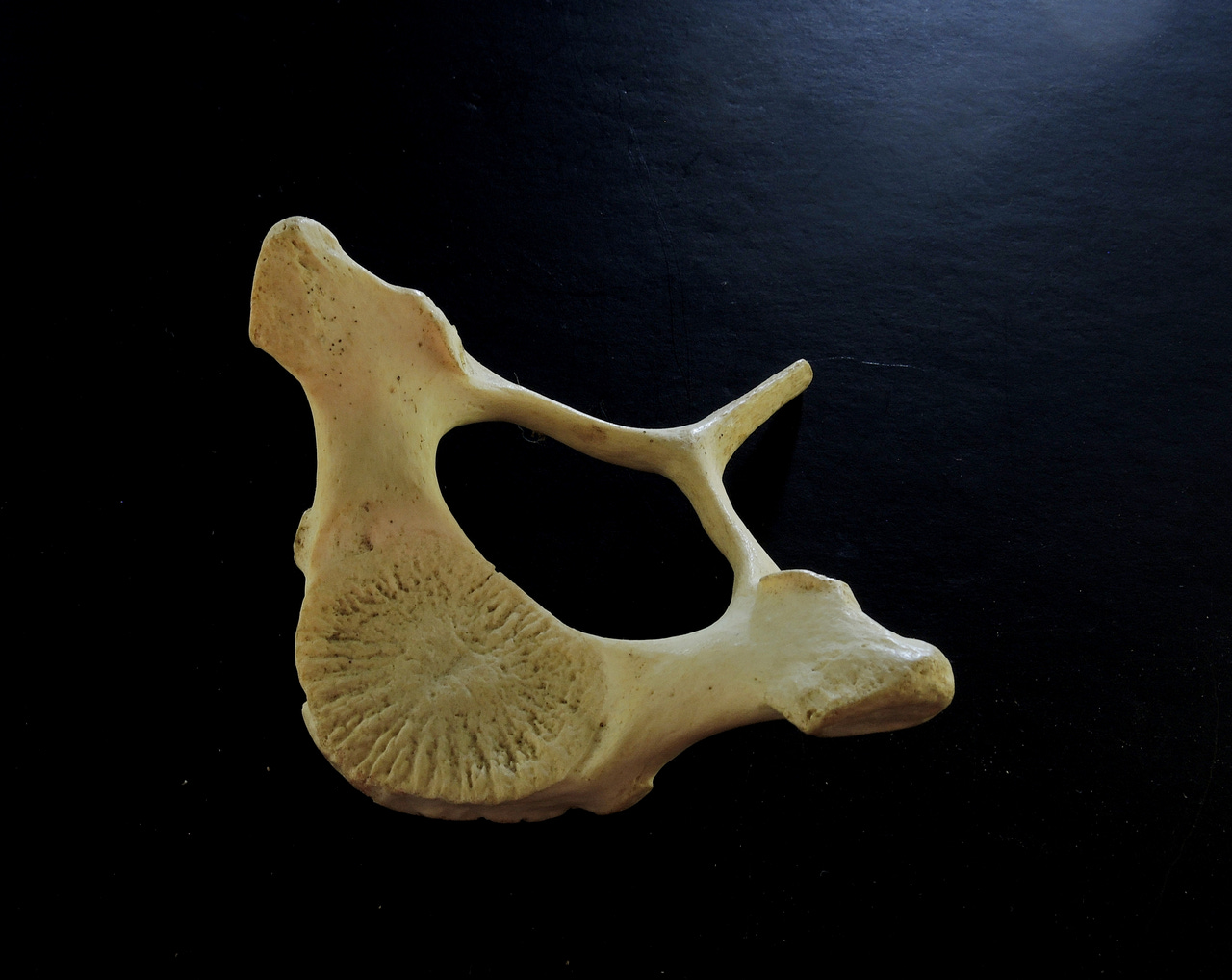
The porpoise whose skeleton I found on the beach back in the 1970s has been a Gift That Keeps on Giving because it provided me with several interesting Found Objects. I guess you would expect that from a complete skeleton--- skull, flippers, ribs, vertebrae, the whole shootin’ match. I posted an essay on the porpoise’s neck, or what little there is to it. But my focus here is that this animal died young, its life cut short by an unknown agent. How do I know this? The bones told me so, and the evidence is in the image below.
The clue derives from the fact that mammals have determinate growth, that is, they grow to a characteristic size and then stop. This contrasts with many animals such as fish, lobsters, silverfish, and most plants, all of which grow indefinitely (indeterminate growth). Humans are an obvious example of determinate growth: we stop growing in early adulthood (at least in height, sigh). There are a lot of theories about why some creatures grow indefinitely and others don’t, but that is a topic for a more theoretically inclined essayist.

As in all kinds of growth, organs of the animals grow in concert, although they may not all grow at the same rate. For soft tissues there is no conflict between growth and function, but for growing bones that provide skeletal support, articulation (movable joints) conflicts to some degree with growth in length. Growing in girth isn’t much problem--- the bone is encased in a living tissue that deposits layers of bone on the outside of the existing bone and thickens it. Length is more of a problem because where the bones articulate with one another one bone moves and slides over another. If length growth were dependent on the same living tissue that adds girth on the bone’s exterior, then sensitive tissues would grind against each other, and that will not do!
Mammalian bone is first laid down as cartilage in the embryo and then ossified into bone, but a thin layer just short of the articular surfaces is not ossified. This layer, or growth plate, separates the main body of the bone (in long bone, the diaphysis) from its articulating end (the epiphysis). So, no live cells are harmed in this video of flexing joints. The growth plate contains stem cells that develop into the full range of bone tissues --- bone cells, blood-producing cells, and marrow cells--- and adds bone cells where it contacts the growing end of the main bone and the epiphysis. These are then ossified, thereby lengthening the bone without interfering with moving surfaces. As long as the animal is growing, the growth plates remain un-ossified, pumping out the cells that make the bone grow.
But of course, when mammals stop growing, their bones also no longer grow, and the growth plates become superfluous. The growth plate then becomes ossified to fuse with the diaphysis and epiphysis, forming a single, solid bone. If a mammal dies young before this happens, the growth plate rots (not being ossified) so that the main bone body separates from the articular surface. In my porpoise, the separation of these articular discs from the main part of the vertebrae (and other bones) told the story of its youthful death. Most of the other bones told the same story. Even the epiphyses of the ribs had separated (and were lost).

Many a TV crime serial features brilliant forensic scientists that provide the detective with evidence of the how’s and when’s of the victim’s death. But really, you don’t have to be a forensic scientist to see that the mammal whose bones you are viewing died young. Anybody can tell.


Who knew that this fire ant specialist was an orthopedic surgeon. A terrific explanation. After digestion of this growth information, one can appreciate how a century or more ago when vitamin D deficiency was a widespread problem resulting in weakness at growth plate locations, bowing of bones during youth (couldn't stop a pig in a ditch) was more common. Now if there is a very undesirable shortening of one leg due to a fracture (less common now) there are growth charts which can be consulted and combining age along with width (thickness of growth plates demonstrated on imaging) one can surgically damage the growth plate on the longer side in those calculated to be sufficiently tall at maturity, to attempt leg length equalization. The Vitamin D deficient rickets along with a genetic condition of Vitamin D resistant rickets, is an unlikely cause of lower limbs bowing in developed countries. Most commonly seen bow legs these days are due to abrading of the inner side of the joint after maturity due to wear and tear arthritis.